baseline vaginal ultrasound
People might experience some light discomfort during it but this should go away afterward. Before initiating tamoxifen baseline vaginal ultrasound sonohysterography or office hysteroscopy to exclude the presence of endometrial polyps appears appropriate.

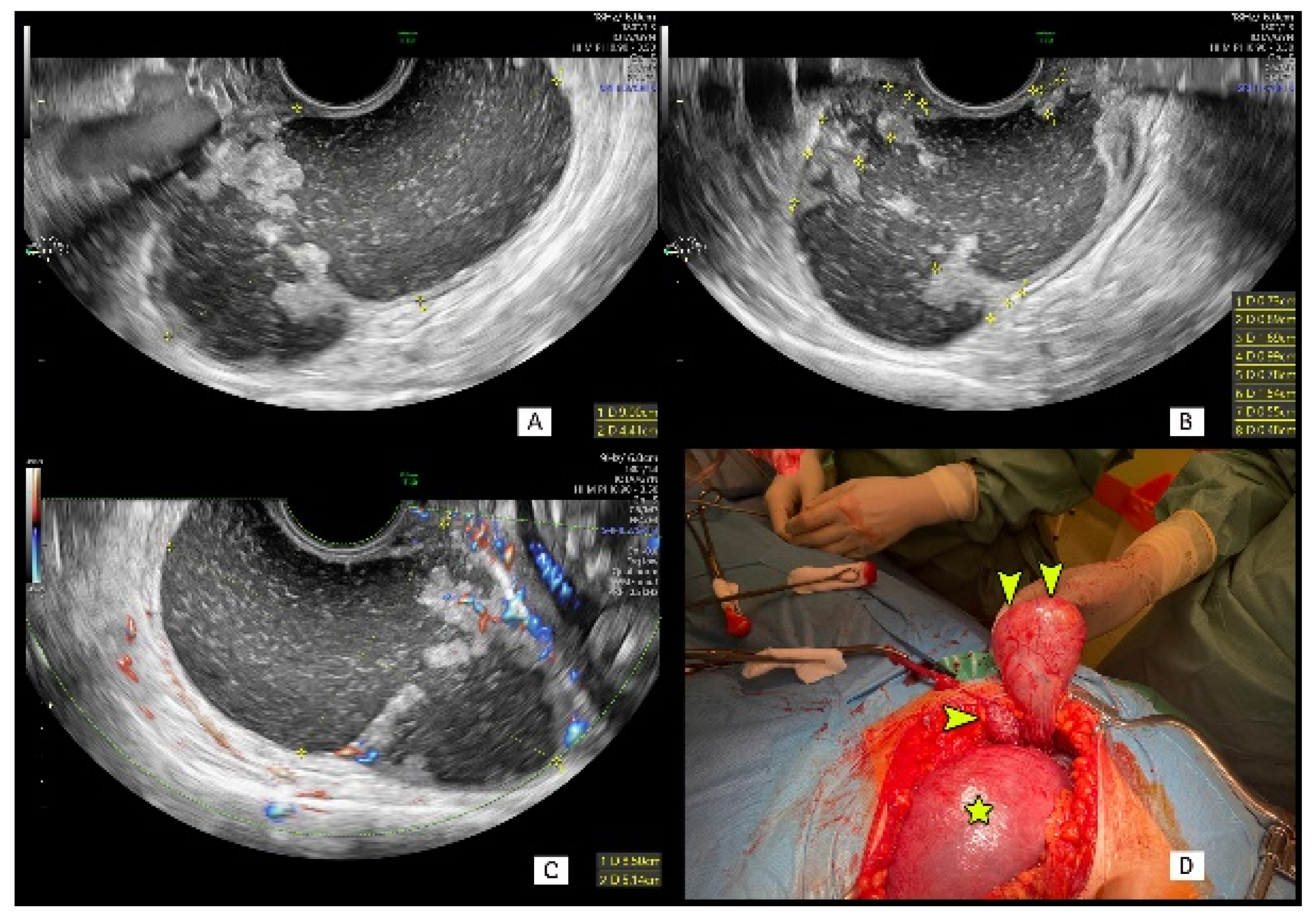
Transvaginal Ultrasound Guided Biopsy Of Deep Pelvic Masses Plett 2016 Journal Of Ultrasound In Medicine Wiley Online Library
After the ultrasound is complete the vaginal probe will be removed.

. In some cases an endometrial sample or biopsy may be performed. When a doctor wants to see the condition of a womans reproductive organs they will use ultrasound to do a pelvic scan fertility test. It is also used to diagnose pelvic pain menstrual and gynecological problems.
There are two methods of performing pelvic ultrasound. A transvaginal ultrasound gives your fertility care team a look at your reproductive organs including your uterus and ovaries. Some doctors use ultrasound during embryo transfer.
A transvaginal ultrasound is a safe scan with no known risks. If you are or have been on Tamoxifen Armidex Anastrozole or Femara it is important that you get a baseline ultrasound of your uterus and then follow up with a second ultrasound two years later. If you discover vaginal bleeding after the transfer it does not mean that the procedure was unsuccessful.
Your doctor might order this test to diagnose a condition or to check. When you are a new patient we often start with a transvaginal. The ultrasound scanner has a probe that gives off sound waves.
A pelvic ultrasound allows quick visualization of the female pelvic organs and structures including the uterus cervix vagina fallopian tubes and ovaries. A pelvic ultrasound is a test that uses sound waves to make pictures of the organs inside your pelvis. This is known as your baseline ultrasound.
Because symptoms may come and go. Before I started these drugs my first ultrasound 2 years ago showed my uterus to be in perfect condition. Female Complete Pelvic Ultrasound Protocol.
This assesses the overall state of your uterus ovaries and fallopian tubes the tubes that connect the ovaries to the uterus. Bleeding is also more common following IVF. Ovarian stimulation and monitoring.
For ultrasound TVS evaluation it is important that the probe once introduced into the vagina is gradually moved upwards while simultaneously evaluating the anterior and posterior vaginal walls. Baseline or screening ultrasound to assess the pelvic anatomy of the uterus including uterine lining and bilateral ovaries. An ultrasound scan is a procedure that uses high frequency sound waves to create a picture of a part of the inside of your body.
It is may also be used during pregnancy to monitor the health and development of the embryo or fetus. Final maturation HCG trigger shot and oocyte retrieval. The number of antral follicles varies from month to month.
Pelvic ultrasound but follow-up recommendations from our ultrasound section varied widely with frequent recommendations for follow-up of. However many studies have not shown that supplementation with estrogen for thin. Different compared to baseline 23 v 22 p070 However follow-up was recommended in 16 patients 5 of total significantly decreased compared to baseline 12 v 5 p0004.
A pelvic ultrasound is a noninvasive diagnostic exam that produces images that are used to assess organs and structures within the female pelvis. The purpose is to check that there are no unusual cysts on the ovaries before starting the fertility drugs. Oral andor vaginal Ethinyl estradiol or transdermal estradiol can be given during the CC cycle to increase the endometrial thickness.
Ultrasound uses a transducer that sends out. The ultrasound may take 1530 minutes. Ultrasound is also used during IVF for egg retrieval to guide the needle through the vaginal wall to the ovaries.
The posterior vaginal wall is in close proximity to the anterior. A thin catheter tube will then be inserted into your cervical canal to enter the uterine cavity. Pelvic scans are considered to be very safe and there are no risks associated.
The Basal Antral Follicle Count along with the womans age and Cycle Day 3 hormone levels are used as indicators for estimating ovarian reserve and the womans chances for pregnancy with. The vagina is limited on its outer surface by the hyperechoic visceral facia. A Pelvic ultrasound scan is the most effective imaging modality used to examine the uterus and ovaries.
Your doctor will insert a speculum just like for a Pap test and cleanse the cervix with a betadyne soap solution. The follicles can be seen measured and counted on Cycle Days 2 3 and 5 by using ultrasound. In males a pelvic ultrasound usually focuses on the bladder and prostate gland.
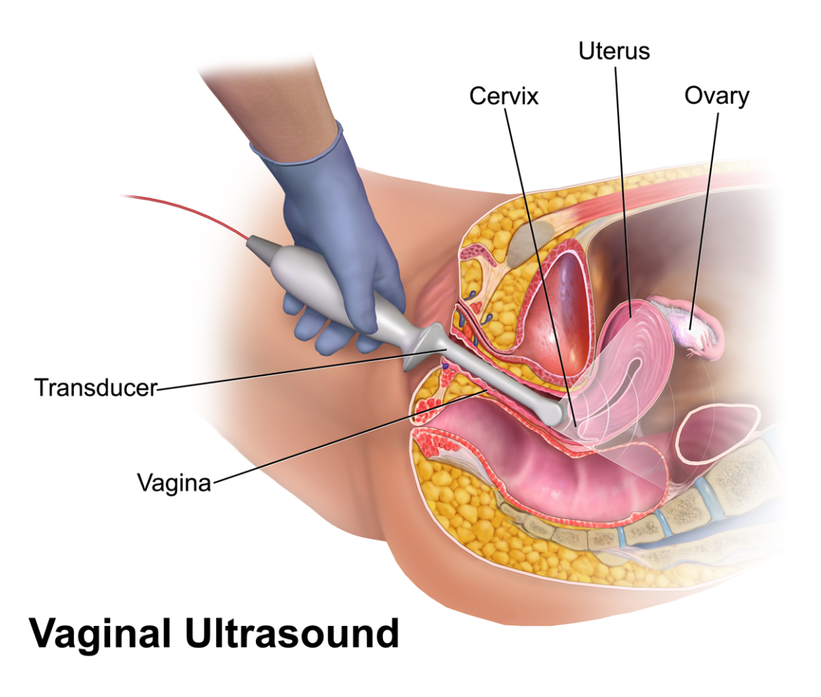
We will you ask you to get a blood. Transvaginal meaning across or through the vagina ultrasound uses sound waves to create images of the vagina cervix uterus and ovaries. The probe looks a bit like a microphone.
The sound waves bounce off the organs inside your body and the probe picks them up. A transvaginal ultrasound can serve many purposes so the goal of yours depends on the type of ultrasound our Las Vegas fertility doctors prescribe. Supra-pubic through a full bladder.

Transvaginal Ultrasound Examination Of The Endometrium In Postmenopausal Women Without Vaginal Bleeding Jokubkiene 2016 Ultrasound In Obstetrics Gynecology Wiley Online Library

Transvaginal Ultrasound Images Showing Typical Features Of A Right Download Scientific Diagram

Transvaginal Ultrasound Guided Biopsy Of Deep Pelvic Masses Plett 2016 Journal Of Ultrasound In Medicine Wiley Online Library

Diagnostics Free Full Text Sonographic Assessment Of Complex Ultrasound Morphology Adnexal Tumors In Pregnant Women With The Use Of Iota Simple Rules Risk And Adnex Scoring Systems Html
Gynaecology Ultrasound Women S Ultrasound Melbourne

Transvaginal Ultrasound Youtube

Sonographic Characterization And Surveillance Of Paravaginal Smooth Muscle Tumor Of Uncertain Malignant Potential Zamora Journal Of Clinical Ultrasound Wiley Online Library

Advanced Pelvic Ultrasound In House At Veritas Fertility Surgery

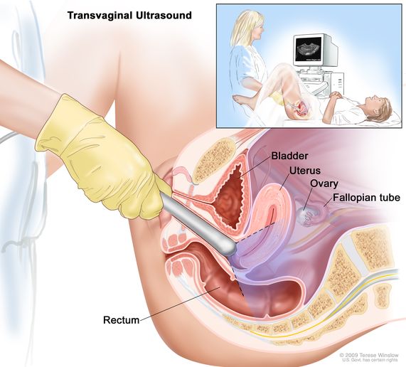
Definition Of Transvaginal Ultrasound Nci Dictionary Of Cancer Terms Nci

Serial Transvaginal Ultrasound Images Evaluated Follicle Number Per Download Scientific Diagram
Transvaginal Ultrasound Showing A Thick Endometrial Cavity With Download Scientific Diagram

Transvaginal Ultrasound Guided Biopsy Of Deep Pelvic Masses Plett 2016 Journal Of Ultrasound In Medicine Wiley Online Library

Advanced Pelvic Ultrasound In House At Veritas Fertility Surgery

Distribution Of The Studied Sample By Transvaginal Ultrasound Findings Download Scientific Diagram

Transvaginal Ultrasound From A 28 Year Old Woman With A Left Ectopic Download Scientific Diagram


Comments
Post a Comment